Case Report | DOI: https://doi.org/CCSRR-CR-26-46
A Silent Obstruction by Gallstones: An Atypical Case of Painless Biliary Blockage at The Duodenal Papilla
Abstract
Introduction: Gallstone disease usually presents with biliary colic or other painful symptoms caused by obstruction of the biliary tree. Painless obstruction of the duodenal papilla by migrated gallstones is rare and may delay diagnosis.
Case presentation: A 60-year-old European female with a several-year history of incidentally diagnosed gallstones remained completely asymptomatic, with no episodes of biliary colic, jaundice, or digestive complaints. Routine follow-up abdominal ultrasound revealed multiple gallstones in the gallbladder and additional stones at the duodenal papilla and proximal duodenum, suggesting migration and partial biliary obstruction. Liver function tests showed cholestatic changes, but the patient reported no pain or systemic symptoms. Endoscopic retrograde cholangiopancreatography (ERCP) was performed, with successful extraction of the papillary stones. An elective laparoscopic cholecystectomy was subsequently planned to prevent recurrence and possible complications.
Conclusions: This case illustrates that significant obstruction at the duodenal papilla can occur without pain, challenging the conventional expectation that biliary obstruction is invariably symptomatic. Gradual onset of obstruction, altered visceral pain pathways, autonomic neuropathy, and potential bilioenteric decompression may contribute to silent presentations. Awareness of such atypical cases is essential, and regular imaging follow-up in patients with known cholelithiasis can facilitate early detection and timely intervention before serious complications develop.
Abbreviations:
CBD : common bile duct
ERCP : endoscopic retrograde cholangiopancreatography
LFTs : liver function tests
MRCP : magnetic resonance cholangiopancreatography
EUS : endoscopic ultrasonography
Introduction:
Gallstones are solid crystalline deposits that form within the biliary tract, most commonly in the gallbladder, and are composed primarily of cholesterol, calcium salts, and bilirubin (1,2). Cholelithiasis is prevalent worldwide and may lead to biliary obstruction when stones migrate into the common bile duct (CBD) or lodge at the duodenal papilla (1,2). Obstruction typically results in acute right upper quadrant or epigastric pain, often associated with nausea, vomiting, and sometimes jaundice or pancreatitis (3–5).
In most patients, biliary obstruction is painful due to increased intraductal pressure, distension of the bile ducts or gallbladder, and inflammation activating visceral nociceptors (3,5). However, there are rare situations in which significant obstruction occurs with minimal or absent pain, contradicting traditional clinical expectations (3). Such presentations are diagnostically challenging and may lead to delayed recognition and treatment, despite the potential for serious complications including acute cholangitis, pancreatitis, or secondary biliary cirrhosis (5).
This report describes a patient with long-standing cholelithiasis in whom multiple stones migrated to the duodenal papilla and proximal duodenum, causing biliary obstruction without pain or other typical symptoms. The possible pathophysiological mechanisms underlying this painless presentation are discussed, together with the diagnostic approach and management strategy.
Case presentation:
A 60-year-old European female was followed in the outpatient setting with a known history of gallstones, first identified incidentally during a routine abdominal ultrasound several years earlier (1,2). She had remained completely asymptomatic over this period, denying any history of biliary colic, jaundice, fever, or digestive complaints. Her medical history was otherwise unremarkable, with no prior abdominal surgery.
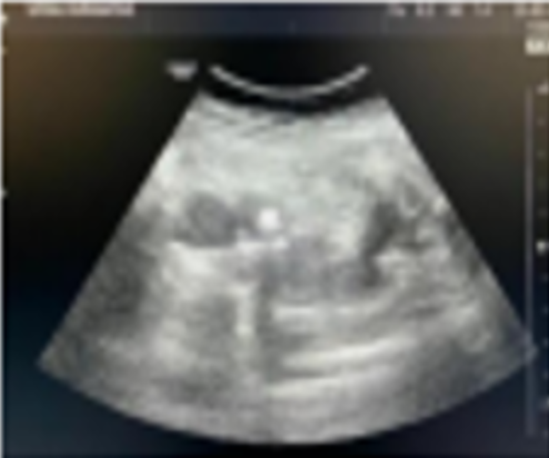
At a scheduled follow-up visit, a general surgeon recommended repeat abdominal ultrasound to reassess the known cholelithiasis. Ultrasound examination demonstrated numerous echogenic calculi with acoustic shadowing within the gallbladder, consistent with multiple gallstones (Figure 1).
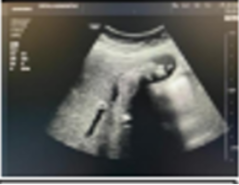
In addition, several stones were visualised at the level of the duodenal papilla (ampulla of Vater) and in the proximal duodenum, indicating stone migration from the gallbladder and biliary tree (Figure 2).The presence of stones distal to the papilla raised the possibility of spontaneous passage into the intestinal lumen.
On physical examination, the patient was afebrile and hemodynamically stable. The abdomen was soft and non-tender, without guarding or rebound, and there was no right upper quadrant or epigastric pain on deep palpation. No palpable masses were noted, and there were no stigmata of chronic liver disease. Laboratory evaluation revealed mild cholestatic abnormalities, including elevated serum bilirubin and alkaline phosphatase, with modest increases in gamma-glutamyl transferase; transaminases were normal to slightly elevated, suggesting at least partial obstruction of the biliary tree in the absence of clinical symptoms (5).
In view of the imaging and biochemical evidence of obstruction, ERCP was performed. Endoscopic inspection of the duodenum identified stones at the major papilla, and cholangiography confirmed filling defects within the distal CBD. A sphincterotomy was carried out, followed by balloon and basket extraction of the stones, with restoration of bile flow (10). No immediate complications occurred, and the patient remained pain free throughout.
After multidisciplinary discussion, an elective laparoscopic cholecystectomy was recommended as definitive management to remove the gallbladder, eliminate the source of gallstones, and reduce the risk of recurrent choledocholithiasis or related complications (11). The patient consented to the procedure, and written informed consent for publication of this case was obtained.
Pathophysiology:
In typical biliary obstruction, pain is the predominant presenting symptom. It is usually described as sharp, colicky, or dull and severe, located in the right upper quadrant or epigastrium, and may radiate to the right shoulder or, less commonly, retrosternally (3). Symptoms are often triggered by fatty meals, can last longer than 30 minutes, and frequently persist for several hours (3). Unlike dyspepsia, acid reflux, or functional gastrointestinal disorders, gallstone-related pain is not relieved by vomiting, bowel movements, or passage of flatus (4).
The mechanisms responsible for pain involve obstruction of bile flow, increased intraductal pressure, distension of the gallbladder and biliary tree, and inflammatory mediators activating visceral afferent fibers that project to the spinal cord and higher centres (3,5). Nevertheless, some patients exhibit biliary obstruction with little or no pain, as in the present case. Several mechanisms may account for this atypical presentation.
One possible explanation is a gradual onset of obstruction, allowing the biliary system to adapt to slowly rising pressures. Progressive dilatation of the CBD can reduce wall tension and dampen afferent nociceptive signaling, leading to minimal symptoms despite significant obstruction (6). Similarly, in chronic pancreatitis, patients may develop CBD or papillary stenosis with only modest changes in liver tests and absent or mild symptoms, reflecting adaptation of the biliary tract over time (6,7).
Altered neural signaling and individual variability in pain perception may also play a role. Structural or functional changes in visceral afferent pathways, central modulation of nociception, or higher pain thresholds could reduce the perception of biliary pain even when obstruction is present (7). Patients with long-standing biliary or pancreatic disease may develop neural remodeling and desensitisation, resulting in atypically mild or absent symptoms (7).
Autonomic neuropathy represents another potential contributor. Autonomic and peripheral neuropathies are common in chronic liver disease and other systemic conditions, and can impair visceral afferent function (8). In such patients, pain related to biliary distension or inflammation may be blunted or absent, leading to silent or oligosymptomatic obstruction (8).
Finally, the presence of a bilioenteric fistula may partially decompress the biliary tree. Cholecystoduodenal or choledochoduodenal fistulas create an abnormal communication between the biliary system and the gastrointestinal tract, allowing bile to bypass the physiological sphincter of Oddi and flow directly into the intestine (9). This reduces intraductal pressure, which may attenuate pain even if stones migrate through the fistulous tract. Surgical repair of the fistula and removal of residual stones are generally recommended to prevent recurrent obstruction and complications, including gallstone ileus (9).
In the present case, the absence of abdominal pain despite papillary obstruction is likely multifactorial, involving gradual obstruction, possible chronic ductal dilation, and individual differences in visceral pain perception (3,6–9).
Additional treatment options in asymptomatic biliary obstruction
ERCP is considered the first-line intervention for choledocholithiasis and papillary obstruction, as it allows both diagnostic cholangiography and therapeutic stone extraction (10). During ERCP, a side-viewing endoscope is advanced into the duodenum, the papilla is cannulated, and contrast medium is injected into the biliary and/or pancreatic ducts to visualise stones, strictures, or leaks under fluoroscopy (10). Sphincterotomy, balloon extraction, basket retrieval, and stent placement can be performed through the same procedure (10).
Laparoscopic cholecystectomy is the standard definitive treatment for gallbladder stones and related complications (11). The minimally invasive approach, performed through several small abdominal incisions, is associated with less postoperative pain, shorter hospital stay, and faster recovery compared with open cholecystectomy (11). Cholecystectomy removes the gallbladder and therefore the source of gallstones, reducing the risk of recurrent biliary colic, choledocholithiasis, cholecystitis, and gallstone pancreatitis (11).
For patients at high surgical risk in whom cholecystectomy is contraindicated or deferred, alternative strategies include endoscopic biliary stenting and pharmacological dissolution therapy with ursodeoxycholic acid (UDCA) (10,12). Endoscopic stents inserted during ERCP can temporarily relieve obstruction due to stones or malignant strictures, but may be complicated by migration, occlusion, or infection, necessitating surveillance and periodic replacement (10). UDCA therapy reduces cholesterol saturation in bile and can gradually dissolve small, radiolucent cholesterol stones in patients with a functioning gallbladder, although recurrence after discontinuation is common and large or calcified stones do not respond well (12).
Discussion:
This case represents an atypical manifestation of biliary lithiasis in which gallstones obstructed the duodenal papilla without causing the classic symptom of biliary pain. In most patients, obstruction at this level would be expected to produce right upper quadrant or epigastric pain due to increased intraductal pressure, gallbladder wall tension, and local inflammation activating visceral nociceptors (3,5). The absence of pain in the present patient therefore warrants consideration of alternative pathophysiological mechanisms (6–9).
Gradual progression of obstruction, chronic ductal dilatation, and neural adaptation may have allowed the biliary system to accommodate elevated pressures without triggering an acute nociceptive response (6,7). Potential contributions from autonomic neuropathy, even in the absence of overt systemic disease, and individual variability in central pain processing should also be acknowledged (7,8). Furthermore, partial or intermittent drainage of bile, whether through a functionally incompetent sphincter of Oddi or an unrecognised bilioenteric fistula, could limit pressure peaks and prevent colicky pain while still permitting biochemical evidence of cholestasis (9).
The clinical implications of asymptomatic biliary obstruction are significant. Silent obstruction may remain undetected until advanced complications occur, including cholangitis, chronic pancreatitis, secondary biliary cirrhosis, or gallstone ileus (5–7). Therefore, clinicians should maintain a high index of suspicion in patients with known cholelithiasis and abnormal liver tests, even in the absence of pain (3,5). Routine imaging with ultrasound, MRCP, or EUS provides a non-invasive means of detecting occult choledocholithiasis or papillary obstruction (5,13). ERCP retains a central role as both a diagnostic and therapeutic modality in such cases (10).
Elective laparoscopic cholecystectomy after clearance of the CBD is widely recommended to prevent recurrent symptoms and complications (11). In carefully selected high-risk patients, endoscopic and medical options can be used to palliate or temporise obstruction, but long-term outcomes may be inferior to definitive surgical management (10–12). This case underscores the importance of not dismissing abnormal imaging or biochemical findings simply because the patient is asymptomatic.
Declarations:
Ethics approval: Not applicable for a single anonymised case report, in accordance with local regulations.
Consent to participate: Not applicable.
Consent for publication: Written informed consent for publication, including clinical data and imaging, was obtained from the patient.
Availability of data and materials: All relevant data are available from the corresponding author on reasonable request.
Competing interests: The authors declare no competing interests.
Funding: No specific funding was received for this work.
Acknowledgements: The authors gratefully acknowledge the contribution of Dr Claudiu Iulian Bădulescu, General Surgery Department, Medlife Humanitas Hospital, Cluj-Napoca, Romania, for his involvement in the surgical management and clinical discussion of this case.
Authors’ contributions: The primary author was responsible for data collection, literature review, manuscript drafting, and manuscript revision. All authors contributed to the clinical management of the patient and approved the final version of the manuscript.
References
-
Stinton LM, Shaffer EA. Epidemiology of gallbladder disease: cholelithiasis and cancer. Gut Liver. 2012;6(2):172–87.
View at Publisher | View at Google Scholar -
Shaffer EA. Gallstone disease: Epidemiology of gallbladder stone disease. Best Pract Res Clin Gastroenterol. 2006;20(6):981–96.
View at Publisher | View at Google Scholar -
Portincasa P, Moschetta A, Petruzzelli M, Palasciano G, Di Ciaula A, Pezzolla A. Gallstone disease: symptoms and diagnosis of gallbladder stones. Best Pract Res Clin Gastroenterol. 2006;20(6):1017–29.
View at Publisher | View at Google Scholar -
National Health Service. Gallstones. NHS; 2017 Oct 20. Available from: https://www.nhs.uk/conditions/gallstones/
View at Publisher | View at Google Scholar -
Del Vecchio Blanco G, Gesuale C, Varanese M, Monteleone G, Paoluzi OA. Idiopathic acute pancreatitis: a review on aetiology and diagnostic work-up. Clin J Gastroenterol. 2019;12(6):511–24.
View at Publisher | View at Google Scholar -
Kalvaria I, Bornman PC, Marks IN, et al. The spectrum and natural history of common bile duct stenosis in chronic alcohol-induced pancreatitis. Ann Surg. 1989;210(5):608–13.
View at Publisher | View at Google Scholar -
Warshaw AL, Schapiro RH, Ferrucci JT, Galdabini JJ. Persistent obstructive jaundice, cholangitis, and biliary cirrhosis due to common bile duct stenosis in chronic pancreatitis. Gastroenterology. 1976;70(3):562–7.
View at Publisher | View at Google Scholar -
Chaudhry V, Corse AM, O'Brien R, Cornblath DR, Klein AS, Thuluvath PJ. Autonomic and peripheral (sensorimotor) neuropathy in chronic liver disease: a clinical and electrophysiologic study. Hepatology. 1999;29(6):1698–703.
View at Publisher | View at Google Scholar -
Glenn F, Reed C, Grafe WR. Biliary enteric fistula. Surg Gynecol Obstet. 1981;153(4):527–31.
View at Publisher | View at Google Scholar -
Adler DG, Baron TH, Davila RE, Egan J, Hirota WK, Leighton JA, et al. ASGE guideline: the role of ERCP in diseases of the biliary tract and the pancreas. Gastrointest Endosc. 2005;62(1):1–8.
View at Publisher | View at Google Scholar -
Abraham S, Rivero HG, Erlikh IV, Griffith LF, Kondamudi VK. Surgical and nonsurgical management of gallstones. Am Fam Physician. 2014;89(10):795–802.
View at Publisher | View at Google Scholar -
Hofmann AF. Medical dissolution of gallstones by oral bile acid therapy. Am J Surg. 1989;158(3):198–204.
View at Publisher | View at Google Scholar -
Romagnuolo J, Bardou M, Rahme E, Joseph L, Reinhold C, Barkun AN. Magnetic resonance cholangiopancreatography: a meta-analysis of test performance in suspected biliary disease. Ann Intern Med. 2003;139(7):547–57.
View at Publisher | View at Google Scholar